top of page

Blood feeding insects that feed on multiple people can spread disease. Mosquitoes are one example where the female mosquito requires a blood meal for protein to properly grow and lay her eggs. This act of blood feeding spreads the malaria parasites to and between people. Another example are sand flies. Sand flies, about ¼ the size of a mosquito, are tiny insect vectors of a different parasite – Leishmania.


Leishmaniasis is the second biggest parasitic killer after malaria. It is estimated to kill around 40,000 people every year, and accounts for about 2.4 million disability-adjusted life-years (DALYs). The Leishmania parasite is transmitted to humans when female sand flies bite humans to obtain blood meals that they require for reproduction. As of August 2022 (the time of writing), there is an outbreak of cutaneous leishmaniasis in Syria near a polluted and drying up river – as reported by VOA news here.

Current treatments of leishmaniasis are highly toxic and/or expensive and no efficacious vaccine exists. Therefore control of this disease requires controlling the Phlebotomine sand flies that transmit Leishmania parasites. A better understanding of sand fly biology and populations is necessary for the control and monitoring of the disease.


Today, we share two chromosome length assemblies for these two sandfly species: Phlebotomus papatasi and Lutzomyia longipalpis. P. papatasi is a vector of the cutaneous leishmaniasis causing parasite Leishmania major in the Old World. L. longipalpis is a vector transmitting Leishmania infantum that causes visceral leishmaniasis in South America.

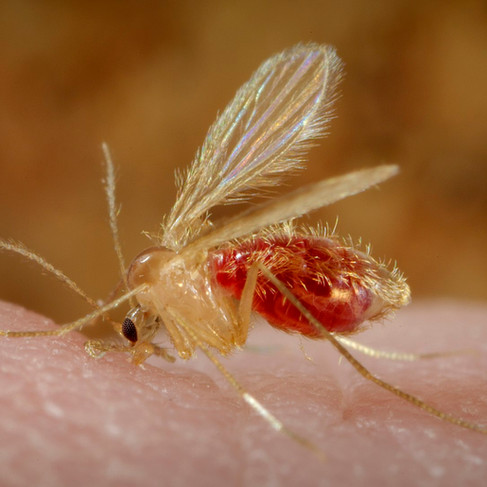
Left: Lutzomyia longipalpis sandfly by Ray Wilson, (2009) PLoS Pathogens Issue Image - Vol. 5(8) August 2009. PLoS Pathog 5(8): ev05.i08. doi:10.1371/image.ppat.v05.i08, [CC BY 4.0]. Right: Phlebotomus papatasi sandfly by Frank Collins, Centers for Disease Control and Prevention's Public Health Image Library (PHIL), identification number #10277, [public domain].

It is our hope that these new high quality reference genomes will enable new and improve ongoing methods of integrated vector control. The improved genome assemblies should enable complex genetic analyses, including descriptions of species complexes, estimation of effective population sizes, monitoring of pesticide resistance allele emergence, and hopefully eventually lead to new leishmaniasis-control methods. Recently, new mosquito genomic resources enabled new vector control methods such as CRISPR-Cas9-based manipulation that create gene drives to prevent malaria transmission and microbiome manipulation with Wolbachia to prevent transmission of dengue. We hope that new reference genomes will enable similar innovative approaches to be developed for sand flies.


The extremely small size of the sand fly made generating these genomes a challenge. Only about 30-50 nanograms of DNA can be isolated from a single male sandfly (males are used here to characterize both the X and the Y sex chromosomes). Using pooled samples and older sequencing technologies in the early sequencing attempts produced extremely fragmented assemblies. We would like to thank researchers at Pacific Biosciences for using these species as a test for their new ultra-low input library generation protocol for the HiFi sequencing technology. When combined with the Hi-C magic performed by the DNA Zoo, we now have highly accurate contiguous sequences scaffolded to chromosome lengths that should stand as a reference for future leishmaniasis prevention research for years to come.


Data from two PacBio Sequel II SMRT cells generated at the BYU sequencing center was assembled with hifiasm (Cheng et al., 2021). Pacific Biosciences generated HiFi data for Phlebotomus papatasi, the draft assembly for was generated by Dr. Sarah Kingan at Pacific Biosciences. For Lutzomyia longipalpis, Sequel II Hifi Data was generated by Oanh Nguyen at the UC Davis Genome Center from a single male using the ultra-low input DNA library kit generated by Dr Kingan and Colleagues at PacBio, and the initial assembly was performed by Stephen Richards using hifiiasm. These high-quality drafts were upgraded to chromosome-length assemblies using Hi-C data from multiple male individuals from the colonies at The University of Notre Dame maintained by Drs. Mary Ann McDowell and Douglas Shoue.


Check out the interactive Juicebox.js instance below for a contact map of five P. papatasi and four L. longipalpis chromosomes, and visit the assembly pages (here and here) for more information and details on the procedure. The genome assemblies are also available on NCBI as Ppap_2.0 (P. papatasi) and ASM2433408v1 (L. longipalpis).


Blog post by Stephen Richards and Mary Ann McDowell.


The native Australian rodent the smoky mouse Pseudomys fumeus, also known by the First People’s name ‘Konoom’ (the name is from the Wadawurrung people of Victoria), was already fighting off extinction when the 2019-2020 fire season hit. The bushfires torched 13.6 million acres, killing an estimated one billion animals and putting more than 100 threatened species at risk. More than 90 percent of the mouse’s habitat was razed by the conflagration. The fires even came for the mice in captive breeding facilities: nine mice died by smoke inhalation at a captive breeding facility near Canberra in February 2020.

ree
The smoky mouse, Pseudomys fumeus. Photo by David Paul, Museums Victoria [CC-BY 4.0]

The smoky mouse is a gentle little mouse with a two-toned pink and grey tail and very soft blue-grey fur which gives them their name. It was described from two specimens first collected in the Otway Ranges, Victoria, in 1933; specimens were later found in the Grampians in 1963, Gippsland in 1971, the Australian Capital Territory (ACT) in 1985 and in New South Wales (NSW) in 1993.


Despite once being widespread, populations of the smoky mouse now tend to be small and fragmented. The species has not been seen in the ACT since the initial capture of two individuals in 1985 and 1986. In Victoria, smoky mice were once found in the Otways and Far East Gippsland but since the 1980s have only been recorded in the Grampians, Central Highlands and Alpine regions of the state. In NSW, the species occurs in alpine regions of Kosciusko National Park and southeastern forests near Nullica.


An active recovery plan was established for the species in 2006. As part of this plan, two captive populations have been established from NSW sources. The first releases from these captive populations are happening into southeastern forests and into a predator proof reserve in the ACT.


To support ongoing conservation efforts, DNA Zoo teamed up with Museums Victoria Senior Curator of Mammals Kevin C. Rowe to release the chromosome-length assembly for the species.


The genome draft was generated with short-insert size Illumina reads [500 PE reads] and scaffolded to chromosome length genome with Hi-C [450 PE reads]. See our Methods page for more detail on the procedure.


The chromosomal-length reference genome will be used to map genomes from 70 smoky mouse individuals from across the species distribution range in the Grampians in western Victoria to southeastern New South Whales (see map below). The data will include historical samples from the holotype specimen (1933) and extirpated populations in the Otways, eastern Victoria, parts of the Grampians as well as contemporary samples.

ree
Geographical distribution of smoky mouse resequencing data by Museums Victoria. Samples include specimens from 1933 (the holotype) as well as contemporary samples from to 2020. Inset map shows details of sampling in Grampians National Park. Geographic centroids of sample clusters are indicated with yellow stars. Each circle represents data from an individual smoky mouse. Shading of circles indicate decade of collection from the 1930s to 2010s.

Samples from the Grampians population are of particular interest. The Grampians population is the most isolated, removed by about 350 km from the nearest extant population in the Yarra Ranges of the Central Highlands. Since 2012, Museums Victoria and partners have trapped, marked, and collected samples (ear biopsies and faecal pellets) from over 200 smoky mouse individuals in the Grampians providing the most numerous and continuous record of the species in Victoria. Trapping and wildlife camera surveys at more than 100 sites revealed populations of Smoky Mouse are localised to two areas spanning <10 km of the Victoria Range and Mt William Range, respectively. Stay tuned for more information about this population as well as genetic clues on how it persisted despite drought, invasive predators and significant fire.

ree
Detections and non-detections of Smoky Mouse in the Grampians based on Museums Victoria surveys from 2012-2021.

This work was enabled by resources provided by DNA Zoo Australia, The University of Western Australia (UWA) and DNA Zoo, Aiden Lab at Baylor College of Medicine (BCM) with additional computational resources and support from the Pawsey Supercomputing Centre with funding from the Australian Government and the Government of Western Australia.


Blog post by Parwinder Kaur and Kevin C. Rowe


Updated: Aug 31, 2022

The green anole, Anolis carolinensis, is one of over 400 species in the genus Anolis (anoles) and one of over 11,000 species in the group Squamata (squamates). Squamates (lizards and snakes) are the most speciose groups of terrestrial vertebrates on earth. Native to the south-eastern United States, the green anole is known and loved for its vibrant green color and charismatic behavioral displays: during the summer months, male green anoles can be seen walking up walls using their sticky toe-pads, or elsewhere prominently perched in gardens and foliage, furiously doing pushups and extending a flag-like flap of bubblegum pink skin under their chin called a “dewlap.”

ree
Green anole, photo by Bonnie Kircher

These characteristic displays are directed at any males that have invaded their territory, as well as any receptive females that catch their powder-blue eyes. Though males are most likely to be spotted displaying, females also perform these displays. Male and female anoles can be differentiated by differences in overall adult body size but also by the size of their dewlap, (males being larger in both cases). Though named for their gorgeous green hue, green anoles also change color to a silver-y brown. Likely not a camouflage technique, this behavior may correspond instead with environmental or social cues.


Anoles are particularly well studied because of their interesting evolutionary quirks. The genus is often described in textbooks as an example of convergent evolution, a process in which similar traits evolve in different species as a result of similar environmental conditions and not due to evolution from a shared common ancestor. Convergent evolution of morphology, physiology, and behavior has been well characterized in anoles and this group is an important model for understanding evolutionary processes. Anoles thrive in a lab setting, making it easy to adapt modern scientific tools and techniques for use in this system. Recently, CRISPR genome editing was used for the first time in a closely related species, the brown anole (Anolis sagrei), making it the first non-avian reptile to have genome editing capabilities be made available.

The genome for green anole, Anolis carolinensis, was first published in 2011 by Jessica Alföldi et al., and was the first non-avian reptile genome to be sequenced. Since its publication and re-annotation in 2013, the genome has been used widely for studies in evolution, genetics, and development. With over 600 citations, the original publication of this genome transformed the ability of researchers to study the evolution vertebrates.


Today, we share a few tweaks to the existing green anole assembly (AnoCar2.0) including anchoring suggestions for 4 microchromosomes missing in AnoCar2.0. We also share the Hi-C data generated using a fibroblast cell line from a female anole individual, originally frozen back in 1981! We thank Drs. Asha Multani, Sen Pathak, Richard Behringer, Liesl Nel-Themaat and Arisa Furuta in the Department of Genetics at the MD Anderson Cancer Center for sharing this cell line.


This is the 6th member of the Squamata family we've released here on the DNA Zoo Blog, see others here! Browse the 18 chromosomes of the green anole in the interactive JuiceBox.js session below. Note an inversion polymorphism in HiC_scaffold_1: stay tuned for more data to find out if this is a culturing artifact or a primary sample polymorphism.


Join our mailing list

ARC-Logo-Final-2018-01.png

© 2018-2022 by the Aiden Lab.

bottom of page